December 2014
Precise device guidance is important for interventional procedures in many different clinical elds including fetal medicine, regional anesthesia, interventional pain management, and interventional oncology. Whilst ultrasound is widely used in clinical practice for real-time guidance, the image contrast that it provides can be insufficient for visualizing tissue structures such as blood vessels, nerves, and tumors. This study was centered on the development of a photoacoustic imaging system for interventional procedures that delivered excitation light in the ranges of 750-900 nm and 1150-1300 nm with an optical fiber positioned in a needle cannula. Co-registered B-mode ultrasound images were obtained.
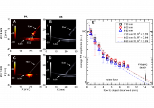
The system, which was based on a commercial ultrasound imaging system, has an axial resolution that is in the vicinity of 100 m and a sub-millimeter, depth-dependent lateral resolution. Using a tissue phantom and 800 nm excitation light, a simulated blood vessel could be visualized at a maximum distance of 15 mm from the needle tip. Spectroscopic contrast for hemoglobin and lipids was observed with ex vivo tissue samples, with photoacoustic signal maxima consistent with the respective optical absorption

distance between the distal end of the ber optic and the polymer tube lled with India
ink, d, varying from 1 to 10 mm. Examples are shown for d = 5 mm (A, B) and for 1 mm
(C, D). Average photoacoustic amplitudes (SROI) in a 4 mm 4 mm region of interest
indicated in (A) for wavelength 750 nm, 800 nm and 850 nm are plotted for d from 1 to
10 mm in (E). The data in (E) represent the average values from 15 image frames; the
error bars represent the standard deviations. Equation (4) is tted to the measured SROI
values for each wavelength and compared with the measured data. The intersections of the tted curves and the noise
oor indicate a maximum imaging depth of around 15
mm for the detection of blood vessels.
spectra. The potential for further optimization of the system is discussed.
Wenfeng Xia
