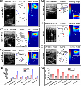
ventions in a sheep in vivo, including: (a) amniotic fluid; (b) muscle femur; (c) right ventricle;
(d) trachea; (e) umblical vein; and (f) stomach; For each insertion, the ultrasound image and the corresponding outlines is on the left, with the tracking image on the right. The signal-to-noise
ratios (SNRs) and the full width at half maximum (FWHM) values of the lateral and axial proles
for tracking images with different excitation methods (single cycle bipolar wave, Barker coding and Golay coding) are compared in (g) and (h) respectively. The dimensions of inserts in all tracking
images are 2 mm 2 mm. Needle tip positions are marked as red crosses according to the positions
of maximums in the tracking images. N: Needle; NT: Needle tip; Sk: Skin; UW: Uterine wall; P:
Placentome; AC: Amniotic cavity; Mu: Muscle; F: Femur; AF: Amniotic
uid; CW: Chest wall;
RV: Right ventricle; LV: Left ventricle; R: Ribs; H: Heart; TV: Tricuspid valve; MV: Mitral valve;
T: Trachea; AW: Abdominal wall; L: Liver; UV: Umbilical vein; M: Membrane; A: Abdomen; S:
Stomach; Pe: Peritoneum.
January 2015
Accurate and efficient guidance of medical devices to procedural targets lies at the heart of interventional procedures. Ultrasound imaging is commonly used for device guidance, however, determining the location of the device tip can be difficult. Various methods have been proposed to improve the visualization of medical devices under ultrasound imaging, a method possessing sufficiently high sensitivity and accuracy, and is fully compatible with the clinical practice is an ongoing pursuit. Ultrasonic tracking is compatible with the current clinical practice and promises to provide high accuracy, but the sensitivity of the method is limited due to the use of miniaturized ultrasound sensors. We present a new generation of the ultrasonic tracking system with dramatically improved sensitivity without sacrificing the accuracy.
A fiber optic hydrophone sensor was integrated into the cannula of a 20 Gauge insertion needle. The sensor which served as an ultrasonic tracker collected transmissions from the ultrasound imaging probe to form an image of the needle tip. Barker and Golay coded excitation were used to improve the sensitivity of the system. The performances of the tracking system in terms of sensitivity and accuracy under different excitation conditions are evaluated in a sheep in vivo in the contexts of brachial plexus nerve blocks and fetal interventions.
During needle insertion performed in the contexts of never blocks and fetal interventions, excellent spatial correspondence was observed between tracking images and ultrasound images. The system has a constantly high accuracy of less than 1 mm. High signal-to-noise ratios (SNRs) were obtained for all tracking images with a maximum of 670 obtained for the insertion in umbilical vein under. Coded excitation signicantly enhanced the SNR of the tracking system compared to bipolar excitation by a factor of around 3 for Barker coding and around 7 for Golay coding. Further image artifacts were observed for tracking images obtained using Barker coded excitation due to the presence range sidelobes. No such artifacts can be observed for Golay coded excitation.
The ultrasonic tracking system using a miniaturized fiber optic hydrophone tracker in combination with coded excitation provided sufficiently high sensitivity and accuracy. The system is compatible with the current clinical practice, and can be used in the contexts of many interventional procedures to improve the accuracy and efficiency to reduced complications.
Wenfeng Xia
