With my colleague Alexander Engels, we have exhaustively reproduced the standard postnatal readouts of this fetal lamb model of open spina bifida.
Harvest was realized at 143 days (term 145 days): after sedation of the ewe, delivery was performed under epidural and local anesthesia by cesarean section so that the lambs were born unsedated and were able survive yet with a foster parent. The ewe was euthanized to allow harvesting of the uterine incision site. The lambs were kept alive for at least 2 days for the readouts as below:
- Morphological examination of the created defect (Meuli-1995): immediately after delivery, macroscopic examination of the surgical site for size and for cerebrospinal fluid (CSF) leakage, detection of fluid leakage surrounding the surgical site with blotting paper.
- Clinical evaluation for neurological deficit (standardized veterinary protocol) (Meuli-1995): general health status, mental status; neurological function including cranial nerves, spinal reflexes, and postural reactions, as well as gait and posture; pain perception of forelimbs, hind limbs, face, and rump assessed by pricking with a needle (superficial pain) and by pinching with a hemostat (deep pain); observation for urinary and fecal incontinence or micturition and defecation problems.
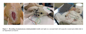
- Measurement of limbs somatosensory evoked potentials (SSEP) under general anesthesia to objectively show sensory transmission to the brain and motor transmission to the distal muscles (Fig.1) (Yingling-1999). SSEP were recorded to stimulation of both posterior tibial and ulnar nerves over the sensory cortex (fig.1).
Figure 1 – Recording of somatosensory evoked potentials (middle and right) on a neonatal lamb with surgically created spina bifida (left) in our department of research 
