With my colleague Alexander Engels, we are improving the readouts of the fetal lamb model of open spina bifida:
- Analysis of watertightness of lesion: just prior to delivery an amniocentesis was performed to test for AcetylCholinesterase and alpha-FetoProtein in the amniotic fluid (should be negative with complete closure): unfortunately the results were not conclusive as these human markers could not be measured in the amniotic fluid.
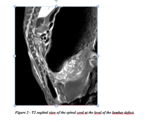
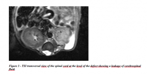
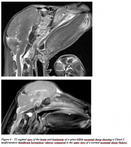
- Brain and spine MRI under general anesthesia using the 3-Tesla SIEMENS MAGNETOM TrioTim syngo MR B17 machine. We analyzed anatomical (T1, T2 and T2 flair) and watertightness (T2-Turbo Inversed Recovery (TIR) sequences) of SB (fig.2-4).
- Histological examination of the operated lumbar spine and of the brain (Meuli-1995): after euthanasia of the lambs, the whole blood was removed via incision of the inferior vena cava. Then we performed a whole-body vascular rinsing with 3L of saline through the abdominal aorta and whole-body vascular fixation with infusion of 3L of the 4% paraformaldehyde. Subsequently the entire spine and the brain were harvested en bloc and fixed for a month in a container full of 4% paraformaldehyde. Serial sagittal and cross sections of the spina and the brain are being prepared and stained with Hematoxylin-Eosin and Gomori’s trichrome (Meuli, Meuli-Simmen et al. 1995).
Figure 2 – T2 sagittal view of the spinal cord at the level of the lumbar defect.
Figure 3 – TSI transversal view of the spinal cord at the level of the defect showing a leakage of cerebrospinal fluid.
Figure 4 – T2 sagittal view of the brain and brainstem of a spina bifida neonatal sheep showing a Chiari 2 malformation (hindbrain herniation) (above) compared to the same view of a normal neonatal sheep (below).
fetal sheep model of spina bifida-May 2015


