Guided Instrumentation for Fetal Therapy and Surgery
Xia Wenfeng
Dr Wenfeng Xia
UCL Postdoctoral Research Associate
Wenfeng received a BSc in Electrical Engineering from Shanghai Jiao-Tong University, in Shanghai, China, in 2005 and an MSc in Medical Physics from University of Heidelberg, in Heidelberg, Germany, in 2007. His graduation project was performed in the Mannheim Biomedical Engineering Laboratories, in Mannheim Germany, focusing on the feasibility study of dental caries removal by picosecond and femtosecond laser ablation. After that, he was an Assistant Researcher in the Institute of Biomedical Optics, Luebeck, Germany, in a project on monitoring and automate control of the therapeutic temperature during retinal photocoagulation utilizing a photoacoustic temperature probe. From 2009 to 2013, he was a PhD student in the Biomedical Photonics Imaging group, University of Twente, Enschede, the Netherlands, developing a 3D photoacoustic mammography laboratory prototype system based on a computed tomography configuration. After receiving a PhD in 2013, he spent one year as a Postdoctoral Researcher to convert the laboratory prototype to a clinical version. He is currently a Postdoctoral Research Associate in the Photoacoustic Imaging Group at UCL, working on interventional photoacoustic imaging and ultrasound devices tracking.
GIFT-Surg focus
Precise image guidance is of great importance for minimally invasive fetal surgeries such as those performed to treat twin-to-twin transfusion syndrome (TTTS). Currently, therapeutic interventions for TTTS
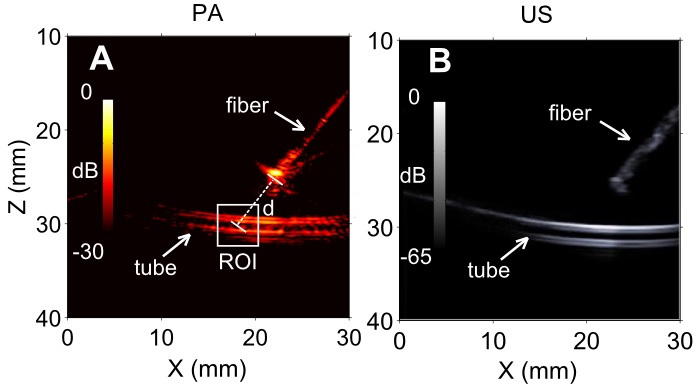
involve the identification of anastomosing vessels on the placenta using a fetoscope, followed by photocoagulation using a continous laser. Inadequate or missing coagulation of the placental anastamoses increases the failure of the procedure. One of the limitations of conventional fetoscopes is that they can provide insufficient sensitivity for vasculature that lies beneath the surface of the placenta due to strong light scattering in biological tissues. Photoacoustic (PA) imaging is a hybrid imaging modality that combines light absorption and ultrasound (US) propagation. It can provide both optical absorption contrast and spectroscopic specificity and subsurface visualisation
of vasculature. It has strong potential to image placental and fetal vasculature with high sensitivity and specificity. In parallel with their developments of photoacoustic imaging, they are developing device tracking systems to precisely track needles that are inserted with ultrasound imaging. With their ultrasonic needle tracking approach, an optical hydrophone is attached to the device to receive transmissions from the transducer array. These received transmissions can be processed to obtain a clear image of the needle tip.
Photoacoustic (left) and ultrasound (right) images of a vessel phantom, with excitation light provided by a fibre adjacent to the vessel (tube).

 UCL Postdoctoral Research Associate